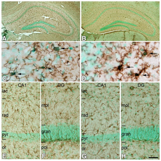
Fig. 4.
Post-mortem time-related changes in microglial and astroglial cells in the hippocampus. a, b Low-magnification photomicrographs showing Iba-1 (microglial cells; a) and GFAP immunoreactivity (astrocytes; b) in immersion-fixed tissue 5 h PT counterstained with methyl green. c, d High-magnification photomicrographs showing that Iba-1-ir microglial cells have larger cell bodies (arrows) and thick, retracted processes in immersion-fixed tissue 5 h PT (d) as compared to perfusion-fixed tissue (c). e–h Low-magnification photomicrographs of CA1 (e, g) and the dentate gyrus (f, h) showing the absence of apparent differences in the patterns of GFAP immunostaining in the brain of animals fixed by immersion after 5 h PT (g, h) compared to animals fixed by perfusion (e, f). DG, dentate gyrus; gran, granular layer; lac, stratum lacunosum moleculare; mol, molecular layer; or, stratum oriens; pol, polymorphic layer; pyr, stratum pyramidale; rad, stratum radiatum. Scale bar (in h): 300 µm in (a, b); 16 µm in (c, d); 40 µm in (e–h)

