Fig. 4.

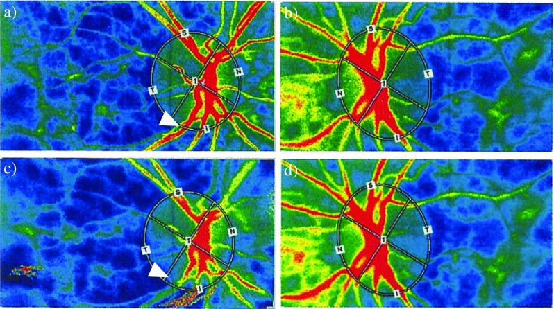
Representative case with diabetic macular edema. Images from a 63-year-old woman with diabetic macular edema in her right eye. Color LSFG map of the right eye (a, c) and left eye (b, d) are shown. Before the IVR in the right eye, the MBR was 17.8 for her right eye (treated eye, a) and 17.8 for the left eye (untreated eye, b). One day after treatment, the MBR decreased to 13.0 (c) in the injected eye and increased slightly to 18.9 in the untreated eye (d). White arrow indicates narrowness of same vessel before and after treatment. LSFG, laser speckle flowgraphy; MBR, mean blur rate
