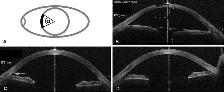
Fig. 1.
OCT measurements. A) Schematic illustration of the TO opening parameter, defined as the angle α over which the TM appeared open in radial OCT sections. B) Illustration of the ACD measurement. The connecting line between the nasal and the temporal angle recess (AR) served as baseline. The maximum distance perpendicular from the baseline to the posterior surface of the cornea was defined as ACD. C) Representative OCT section showing an open TM (white arrow). D) OCT section showing re-closure of Schlemm’s canal by anterior synechia to the TM opening. The scleral spur is located above the white asterisk

