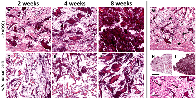
Figure 6.

Time-course of tissue modifications observed by Hematoxylin and Eosin staining in the scaffolds explanted at 2 (A,D), 4 (B,E) or 8 (C,F) weeks after subcutaneous implantation, either with (A–C) or without (D–F) the addition of hADSCs. It is evident that the material was wrapped, and then invaded, by fibroblast-like cells (asterisks in G), and this process was more robust in scaffolds seeded with hADSCs (compare A,B with D,E). Along the observed time-points, the material undergone a visible mineralization that, again, appeared dramatically increased in scaffolds seeded with hADSCs prior to implantation (compare A–C with D–F). In particular, the scaffold appeared almost completely mineralized at 8 weeks (C). The mineralization is evident also comparing the scaffolds sections at low magnification in H (2 weeks, without hADSCs) and I (8 weeks, with hADSCs). The formation of vascular elements is evident in G (arrow) and at higher magnification in J (arrows). Scale bar in A for A-F: 100 μm; in G: 200 μm; in H for H, I: 1 mm; in J: 50 μm.
