
Figure 4.
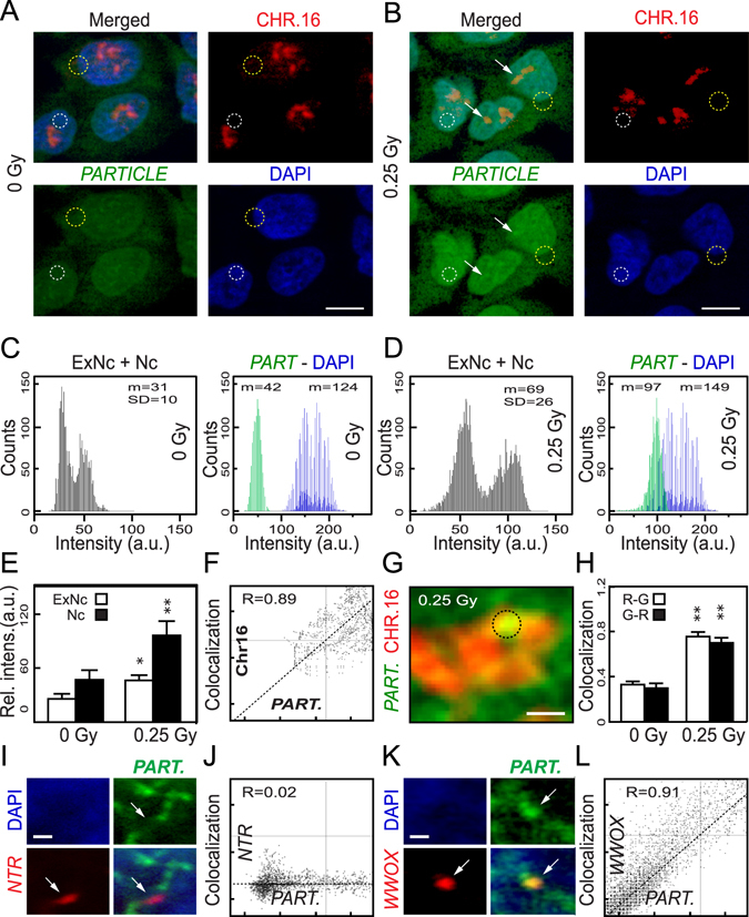
PARTICLE transcripts are predominantly nuclear following irradiation and associated with chromosome 16 harbouring WWOX. (A,B) Representative epifluorescence microscopic images of U2OS sham irradiated (left) or 24 hr post 0.25 Gy (right) labelled with chromosome 16 paint (red: upper) and RNA in-situ hybridisation probes specific for PARTICLE (green: lower). Nuclei stained with DAPI (blue; lower) with merged images (upper). Mean fluorescence intensity analysis determined from regions-of-interest (ROI) indicated by circular dashed lines. Increased intensity over chromosome 16 indicated by arrows. (C,D) Plots of arbitrary units (AU) of overall PARTICLE (PART.) fluorescence intensity in the extra-nuclear (ExNc) plus nuclear (Nc) compartments (left) and intracellular distribution (right; green and blue lines represent PARTICLE and nuclear fluorescence respectively) in sham-irradiated (C) or 24 hr post 0.25 Gy (D). (E) Summary plots illustrating average relative fluorescence intensities pooled from the selected ROIs from the various experimental groups. (F) Scatterplot of arbitrary units (AU) of PARTICLE and chromosome 16 co-localization from a ROI (dashed circle), on (G) a representative high resolution epifluorescence micrograph after chromosomal spreading. (H) Summary co-localization plots for chromosome 16 (ChR = red channel) and PARTICLE (ChG = green channel) in ROIs taken from sham irradiated (0 Gy) or 24 hr post 0.25 Gy. Data are represented as mean ± SEM. (I) Representative epifluorescence microscopic images of U2OS labelled with RNA in-situ hybridisation probes specific for PARTICLE (PART: pseudo green: upper right) and dual labelled probe specific for a predicted Non-Triplex Region (NTR: pseudo red: lower left). Nuclei stained with DAPI (blue; upper left) with merged images (lower right). Arrow indicates absence of PARTICLE at NTR. (J) Scatterplot of arbitrary units (AU) of PARTICLE (PART.) and NTR indicating no co-localization. (K) Representative epifluorescence microscopic images of U2OS labelled with RNA in-situ hybridisation probes specific for PARTICLE (PART: pseudo green: upper right) and dual-labelled probe-specific for WWOX locus (pseudo red: lower left). Nuclei stained with DAPI (blue; upper left) with merged images (lower right). Arrow indicates presence of PARTICLE at WWOX locus. (L) Scatterplot of arbitrary units (AU) of PARTICLE (PART.) and WWOX indicating strong co-localization.