FIGURE 2.

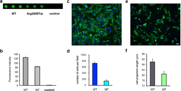
Analysis of the interaction of LAMA5 mutant with SV2A and the ability of the LAMA5 mutant to support cell adhesion and βIII tubulin-positive cell outgrow. (a) Dot blot analysis showing decreased immunoreactivity against SV2A in immobilized lysates from HEK cells transfected with mutant LAMA5 cDNA (wells 4–6) in comparison with lysates from cells transfected with WT LAMA5 cDNA (wells 1–3). (b) Bar graph summarizing measurements of fluorescence intensity in arbitrary units. Control group represents lysates of untransfected cells. (c) Example of normal cell density and neurite outgrowth in a slide coated with WT laminin. (d) Summary bar graph of four transfection experiments showing that cell adhesion to slides coated with laminin-521 containing the LAMA5-Trp2659 was only about 25% of slides coated with the WT laminin-521. (e) Example of low cell density and shorter neurite outgrow in a slide coated with mutant laminin (f) Bar graph showing that βIII tubulin-positive cell projections were overall shorter in slides coated with mutant laminin in comparison with those coated with WT laminin. The calibration mark represents 25 μm in c and e.
