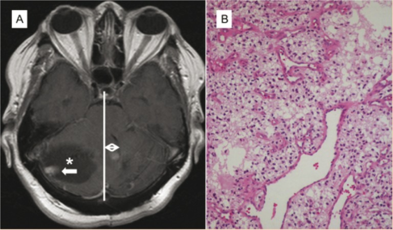
Figure 1.
(A) Axial T1 postcontrast images demonstrate an enhancing mural nodule (white arrow) with accompanying cyst (*) in the right cerebellar hemisphere exerting a mass effect upon the midline and fourth ventricle (double white arrow). (B) Hematoxylin-eosin staining showing scattered large hyperchromatic nuclei, vacuolated cells, and multiple capillaries which are classic features of the cellular type of hemangioblastoma.

