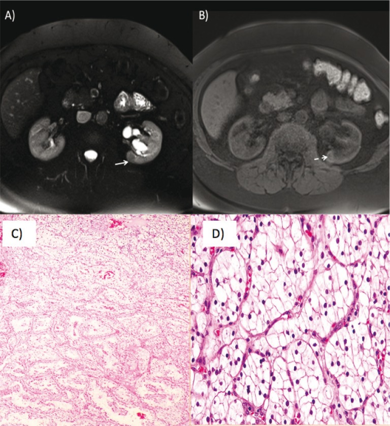
Figure 2.
(A) Axial T2 weighted images show a T2 hypointense lesion in the posteromedial aspect of the midportion of the right kidney (white arrow). (B) Axial T1 postcontrast images show enhancement of the lesion (dashed white arrow) indicating a solid renal mass such as renal cell carcinoma. (C) Low-power- and (D) high-power-hematoxylin-eosin staining showing a typical picture of clear cell renal cell carcinoma with nests of clear cells surrounded by intricately branching vascular septa.

