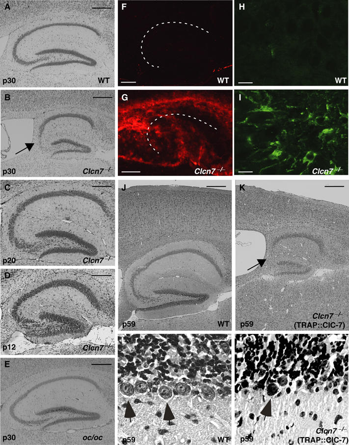
Figure 2.

Neurodegeneration in Clcn7−/−, but not in oc/oc mice. (A–E) Neuronal loss in the hippocampal CA3 region (arrow in B) at p30 as revealed by Nissl staining was found in Clcn7−/− mice (B). oc/oc mice (E) were similar to WT controls (A). At p12, hippocampal sections of Clcn7−/− mice appeared normal (D). A reduction of cell density became visible at p20 (C). These pathological changes were observed in all animals that were analyzed at p30 or older (n>8). Neurodegeneration in Clcn7−/− mice was accompanied by increased staining with the microglial marker GSA (F, G; the pyramidal cell layer is indicated by a dashed line) and for the astrocyte marker protein GFAP (H, I). (K) In 2-months-old Clcn7−/− mice carrying a TRAP::ClC-7 transgene to rescue osteopetrosis, a further degeneration of cerebral cortex and hippocampus (arrow, CA3 region) is visible in Nissl staining as compared to WT (J). Severe loss of Purkinje cells (arrows) in the cerebellum of Clcn7−/− TRAP::ClC-7 mice (M) compared to WT (L) at p59. Scale bars: (A–G, J, K), 0.25 mm; (H, I, L, M), 10 μm.
