
Figure 7.
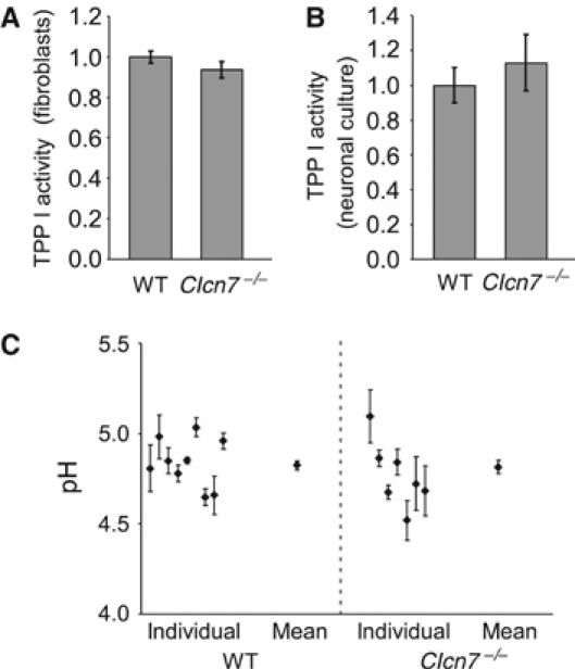
Lysosomal pH and enzyme activity in Clcn7−/− cells. (A, B) Measurement of TPP I activity in living cells using Ala–Ala–Phe–Rhodamine110–Phe– Ala–Ala as a substrate. (A) Intracellular fluorescence generated after a 1 h incubation of fibroblasts with the fluorogenic substrate, as analyzed by FACS. (B) TPP I activity in neuronal cultures, as indicated by fluorescence released into the supernatant and normalized to cell density. (C) Lysosomal pH of WT and Clcn7−/− neurons was determined with dextrane-coupled pH-sensitive dye Oregon green 488 that was loaded by endocytosis. The average pH of WT and KO lysosomes was 4.82±0.03 (s.e.m.) and 4.82±0.04, respectively (n=100 visual fields (containing one to two cells) for WT and 69 for KO; the plot shows pH obtained from individual animals as well as the mean).