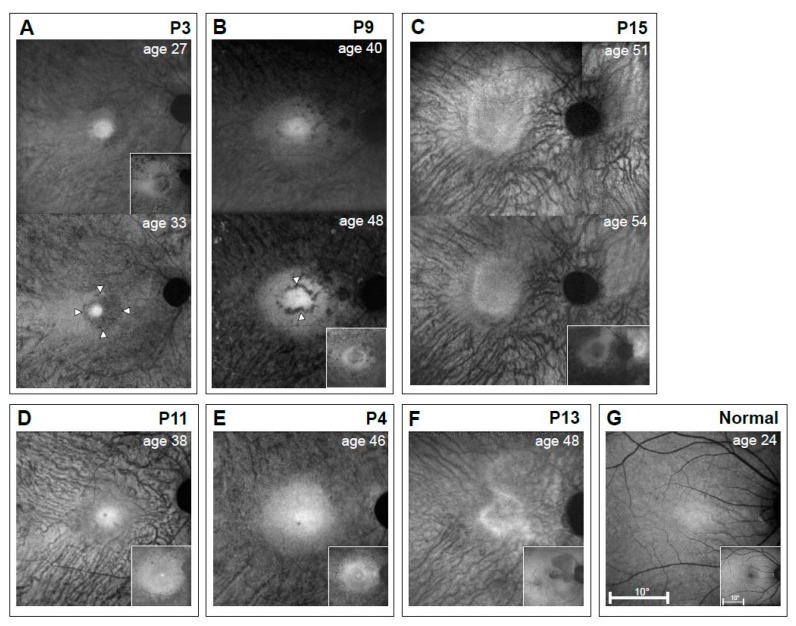
Figure 6.
Autofluorescence imaging results of the macula of a representative normal subject compared with 6 EYS patients. Melanin autofluorescence with Near-infrared reduced-illuminance autofluorescence imaging (NIR-RAFI) is shown in the main panels, and lipofuscin autofluorescence (when available) with short wavelength (SW)-RAFI is shown as insets (lower right panels). (A–C) Serial data from P3, P9, and P15 are over six, eight, and three years, respectively. (D–F) P11, P4, and P13 have images at single timepoints. (G) Images from a 24-year-old normal subject. All eyes are displayed as equivalent right eyes and images are individually contrast stretched for visibility of features. Arrowheads in P3 (age 33) and P9 (age 48) indicate hypo AF rings around the hyper AF central signal.

