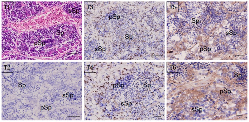
Figure 5.
Localization of HSP60/HSP10 in the mature testis. Immunohistochemical (IHC) positive signals of HSP60/HSP10 immunolabeling are shown in brown. (T1): the whole testis section stained with H&E; (T2): negative control (NC); (T3): different part and developmental phase of testis for IHC with anti-HSP60; (T4): magnify for the IHC with anti-HSP60; (T5): different part and developmental phase of testis for IHC with anti-HSP10; (T6): magnify for the IHC with anti-HSP10, respectively. pSp: primary spermatocytes, sSp: secondary spermatocyte, and Sp: spermatids. Scale bar = 100 um.

