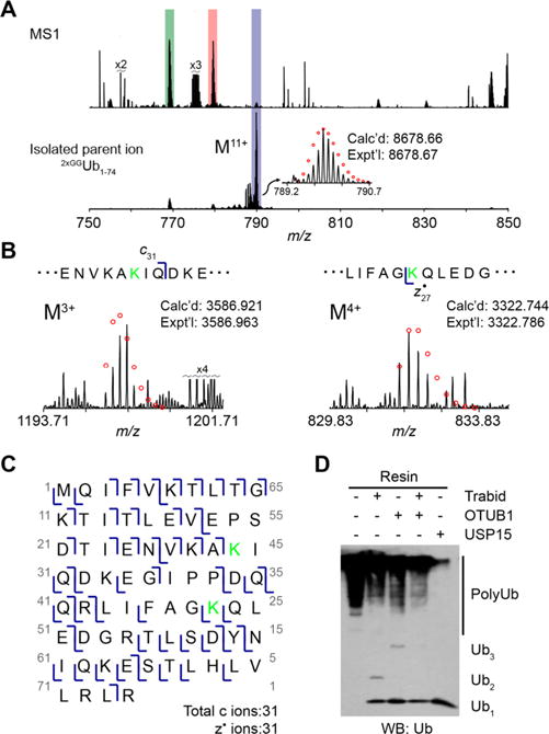
Figure 4.

Characterization of Ub chains isolated with Halo-NZF1 by ETD and DUB restriction analysis uncovering K29/K48 branched chains. (A) ESI MS analysis of the minimally digested Ub chains isolated from HEK cells treated with 10 μM MG132 for 2 h. The spectra correspond to the Ub11+ charge state, where the top spectrum shows all ions present in the mass range and the bottom spectrum shows the isolated 2xGGUb1–74 M11+ parent ion. The red circles represent theoretical isotopic abundance distributions of the isotopomer peaks. calcd, calculated most abundant weight; exptl, experimental most abundant molecular weight. (B) Analysis of ETD fragments showing the presence of a di-Gly modification at K29 and K48. The green K shows the position of a modified lysine. (C) Observed ETD fragments (c and z• ions) mapped onto the sequence of Ub containing a di-Gly modification at K29 and K48. (D) DUB restriction analysis of Ub chains isolated from MG132 treated cells using Halo-NZF1 resin. Aliquots of this resin were incubated with the indicated DUBs overnight at 37 °C. The reaction mixtures were quenched with 6× Laemmli loading dye, separated on a 15% SDS-PAGE gel, and analyzed by Western blot using anti-Ub antibody (P4D1).
