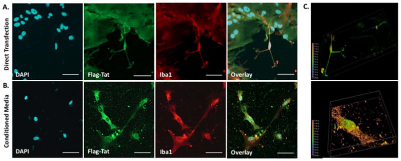
Figure 3. HIV Flag-Tat101 Uptake in Midbrain Microglia.
A. Mouse midbrain microglia in a mixed glia culture at postnatal day 2 were transfected with 1 μg of a plasmid containing the HIV-1 Flag-Tat101 gene (pcDNA3.1+/Tat101-flag(PEV280)), immunostained with the monoclonal ANTI-FLAG® M2 primary antibody (Sigma-Aldrich, St. Louis, MO, USA), polyclonal Iba1 primary antibody (Wako Pure Chemical Industries, Ltd., Richmond, VA, USA), and secondary antibody conjugated to Alexa 488 (Green) or 647 (Red), mounted with DAPI (Blue) Fluoromount-G (SouthernBiotech, Birmingham, AL, USA), and imaged 24 hours post-transfection on a Nikon Imaging System (Nikon A1RMPsi-STORM 4.0, 20X Magnification). N = 6 independent experiments (midbrain of 3–5 mice used for each experiment). B. Another set of mouse midbrain microglia in a mixed glia culture at postnatal day 2 was incubated with conditioned media containing the HIV-1 Flag-Tat protein, immunostained as described above, and imaged 24 hours later (20X Magnification). N = 5 independent experiments (midbrain of 3–5 mice used for each experiment). C. Images represent a depth coded 3D Z-stack of one microglia cell from the 60X magnified image in panel A (top) and panel B (bottom). Scale bar = 50 μm in 20X images and 25 μm in 60X images.

