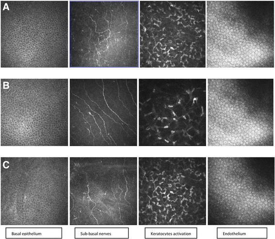
Fig. 1.

In vivo corneal confocal microscopy findings in Tafluprost 0.0015% group, controls and PF Timolol 0.1% group. a Tafluprost 0.0015% group; b Control group; c PF Timolol 0.1% group. The basal epithelium layer images show a significant increase of cell density in therapy groups with respect to control group; no difference appears between Tafluprost and PF Timolol groups. The sub-basal nerve plexus figures show a reduction of the number of nerve fibers with higher tortuosity scores in patients on therapy with respect to controls; Tafluprost and PF timolol groups don’t differ in sub-basal nerve number and morphology. Stromal images underline a significantly higher keratocytes hyperreflective network in therapy groups than in controls. Stromal reflectivity is similar in Tafluprost and PF Timolol groups. The endothelial images show a similar mosaic pattern in the three groups
