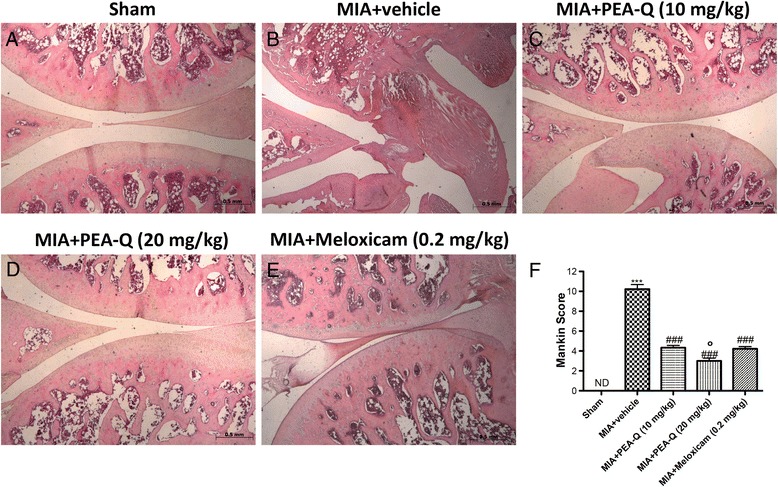
Fig. 5.

Effects of PEA-Q on MIA-induced histological features of OA knee tissue. Histological evaluation was performed by haematoxylin and eosin staining. Panel (a), sham; panel (b), MIA–injected; Panels (c) and (d), PEA-Q treatment; Panels d and (e), meloxicam treatment. Figures are representative of all animals in each group. Panel (f), Mankin score for the various treatment groups. Values are means ± SEM of 10 animals for each group. ***p < 0.0001 vs sham. ### p < 0.0001 vs MIA + vehicle. °p < 0.05 vs MIA + PEA-Q (10 mg/kg) and MIA + meloxicam
