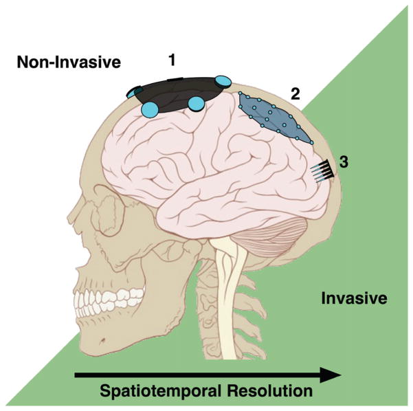
FIG. 1.
Signal resolution and NI placement. In general, the more invasive the NI the higher the accessible spatial and temporal resolution. Scalp-mounted EEG electrodes (1) and ECoG electrodes under the dura (2) record gross cortical oscillations, while intracortical electrode arrays (3) can detect single-cell activity. (Adapted from Lynch & Jaffe, 2006.)

