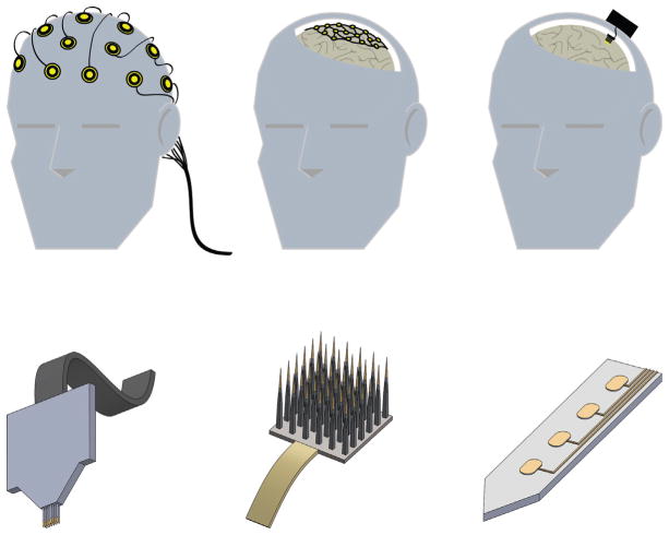
FIG. 2.
Neuroprosthetic interfaces in the CNS. Top: Placement and invasiveness for prominent BMI approaches. Current interfaces interact with the CNS at the scalp (left), the brain surface (middle), or from within the brain (right). Bottom: Examples of intracortical/penetrating neural electrodes. Intracortical NIs may take the form of microwire assemblies (left), arrays (middle), or flat shanks with multiple active recording/stimulation sites (right; center ellipses portray active sites).

