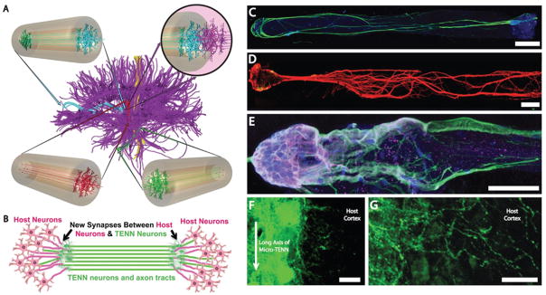
FIG. 4.
Micro-tissue engineered neural networks (Micro-TENNs), consisting of discrete neuronal population(s) with long axonal tracts within a biocompatible micro-column. Micro-TENNs are used for the direct reconstruction of long-distance axonal pathways after CNS degradation. (A) Diffusion tensor imaging representation of the human brain demonstrating the connectome comprised of long-distance axonal tracts connecting functionally distinct regions of the brain. Unidirectional (red, green) micro-TENNs and bidirectional (blue) micro-TENNs can bridge various regions of the brain (blue: corticothalamic pathway, red: nigrostriatal pathway, green: entorhinal cortex to hippocampus pathway) and synapse with host axons (purple; top right). (B) Conceptual representation of a micro-TENN forming local synapses with host neurons to form a new functional relay to replace missing or damaged axonal tracts. (C) Confocal reconstruction of a bidirectional micro-TENN, consisting of two populations of neurons spanned by long axonal tracts within a hydrogel micro-column stained via immunocytochemistry to denote axons (b-tubulin III; green), and cell nuclei (Hoechst; blue). (D) Confocal reconstruction of a unidirectional micro-TENN consisting of a single neuron population (MAP2; green) extending axons (tau; red) longitudinally (adapted from (Cullen et al., 2012)). (E) Confocal reconstruction of a unidirectional micro-TENN, stained via immunocytochemistry to denote neuronal somata/dendrites (MAP2; purple), neuronal somata/axons (tau; green) and cell nuclei (Hoechst; blue). (F) Confocal reconstruction of a transplanted GFP+ micro-TENN showing lateral outgrowth in vivo. (G) Confocal reconstruction showing GFP+ processes extending from a transplanted micro-TENN into the cortex of a rat. Scale bars: 300 μm in C, 250 μm in D, 100 μm in E, 20 μm in F and G. (Reprinted from Struzyna LA, Harris JP, Katiyar KS, Chen HI, Cullen DK, with permission from the Editorial Office of Neural Regeneration Research, Copyright 2015.)

