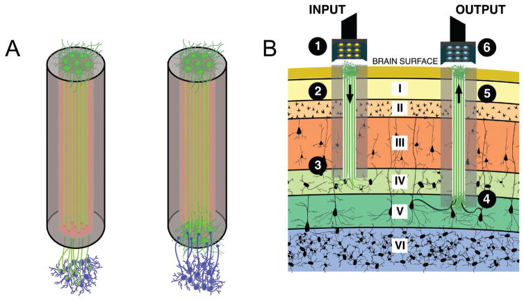
FIG. 5.
Micro-TENNs as “living electrodes.” (A) Unidirectional micro-TENNs (left) synapse with the host (blue cells) and deliver inputs to targeted cortical regions, while bidirectional micro-TENNs (right) may be synapsed by the host and transmit cortical activity. Relevant signal propagation denoted by arrows. (B) Conceptual schematic of the micro-TENNs as “living electrodes” in vivo. Left: Input paradigm. An LED array (1) optically stimulates a unidirectional micro-TENN with channelrhodopsin-positive neurons (2), which synapse with host Layer IV neurons (3). Right: Output paradigm. A microelectrode array (4) records from the neurons of a bidirectional micro-TENN (5), which are synapsed by host neurons from Layer V (6). Roman numerals denote cortical layers.

