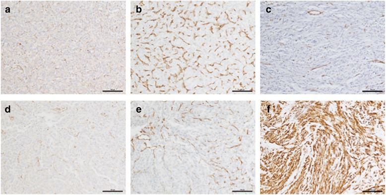
Fig. 3.

Panels a, d SDHB IHC for GIST 1 and GIST 2 shows distinct punctate staining in endothelial cells while tumor cells are negative for SDHB. Panels b, e IHC for endothelial marker CD31 for GIST 1 and GIST 2. Panel c Negative SDHB IHC for a control SDH-deficient RTK-wild type GIST. Panel f Positive SDHB staining for an SDH-competent KIT mutant GIST
