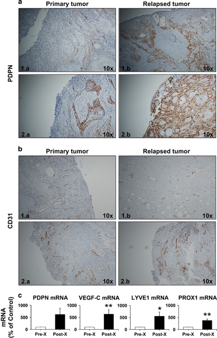
Figure 5.
Evaluation of vascular and lymphatic markers in biopsies from patients diagnosed with HNSCC. Representative images of immunohistochemistry for (a) PDPN and (b) CD31 expression: (1) oral and (2) hypopharyngeal localization; Left panels (1.a, 2.a)—primary tumor; Right panels (1.b, 2.b)—relapsed tumor in the same patient after surgery and chemo-X radiotherapy (brown, PDPN/CD31; blue, hematoxylin - nuclei); (c) quantitative PDPN, VEGF-C, LYVE1 and PROX1 mRNA expression, as percentage of control (0 Gy); * and **, significantly increased values (P<0.05 and P<0.01, respectively) post-, as compared to pre-X radiotherapy.

