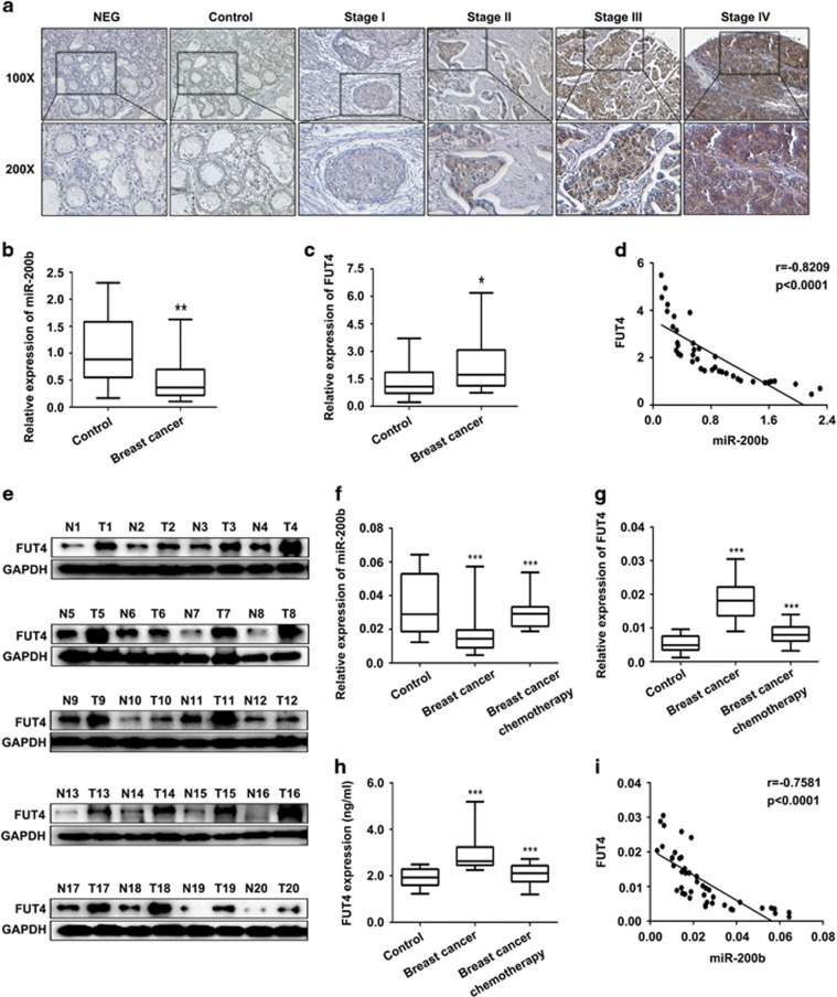
Figure 1.
Low miR-200b and high FUT4 level in breast cancer patient specimens. (a) Detection of FUT4 expression in breast cancer tissue chip (100 cases, pathological grading from stage I to stage IV). Representative images were shown. NEG: PBS was used to replace primary antibody of FUT4. Control: normal human breast tissue. (magnification, × 100 and × 200). (b, c) Real-time PCR for miR-200b and FUT4 levels in tissues of breast cancer (T1-20) and matched adjacent non-cancerous normal tissues (N1-20). (d) The correlation between miR-200b and FUT4 levels in breast cancer and matched normal adjacent tissues. (e) Western blotting analysis for the expression of FUT4 in breast cancer tissues (T1-20) and matched adjacent non-cancerous normal tissues (N1-20). GAPDH served as an internal reference. (f) Expression of miR-200b in serum of healthy controls, breast cancer patients and patients after chemotherapy was detected by real-time PCR. (g, h) Real-time PCR and enzyme-linked immunosorbent assay for the level of FUT4 in serum of healthy controls, breast cancer patients and patients after chemotherapy. (i) The correlation between miR-200b and FUT4 levels in serum of healthy controls and breast cancer patients. *P<0.05, **P<0.01, ***P<0.001.

