Figure 6.
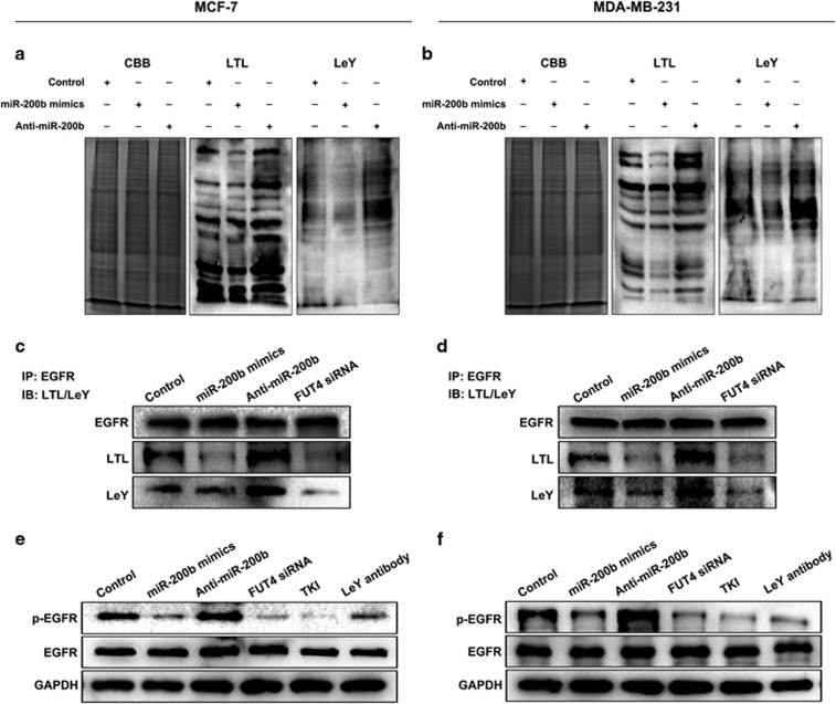
miR-200b decreases α1,3-fucosylation and phosphorylation of EGFR. (a, b) Fucosylation analysis by lectin blotting. Cells were transfected with miR-200b mimics and Anti-miR-200b, respectively. After 48 h culture, protein samples from cell lysates were harvested for detection. LTL lectin were used for detection of α1,3-fucosylation epitope and anti-LeY IgM antibody for assay of specific fucosylated antigen with α1,3-fucosylation linkage. CBB, Coomassie brilliant blue was applied as an equal loading control. (c, d) α1,3-Fucosylation of EGFR. Cells were treated with miR-200b mimics, Anti-miR-200b or FUT4 siRNA. Immunoprecipitation (IP): anti-EGFR antibody pulls down protein. Immune blot (IB): the level of α1,3-fucosylation was detected by LTL lectin and anti-LeY IgM antibody. (e, f) Phosphorylation of EGFR by western blotting. Cells were plated onto six-well plate and treated with miR-200b mimics, Anti-miR-200b, FUT4 siRNA, EGFR activation inhibitor (TKI) and anti-LeY IgM antibody (1:200), respectively. Anti-EGFR antibody (1:500) and anti-phosphorylated EGFR antibody (1:500) were used to detect EGFR activation.

