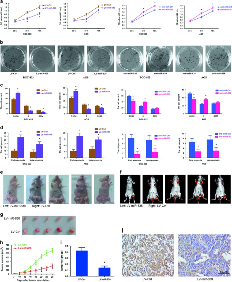
Figure 3.
miR-638 inhibits GC cell proliferation in vitro and in vivo. (a) Cell proliferation was examined by MTT assay at 24, 48 and 72 h after transfection with LV-miR-638 or anti-miR-638. *P<0.01. (b) Cell colonies were analyzed by colony formation assay 12 days after transfection. (c) Detection of cell cycle by flow cytometry analysis was visualized using propidium iodide staining. Histograms showed the percentage of cells in the G1/G0, S and G2/M phases. *P<0.01. (d) Detection of cell apoptosis by flow cytometry analysis was visualized using Annexin-V/propidium iodide staining. The data showed the percentage of early- and late-apoptotic cells. *P<0.01. (e) Gross morphology of tumors injected with either LV-miR-638 or LV-Ctrl cells after 28 days. (f) Small animal imaging analysis was used to assess tumor volume in situ at day 28 during tumor development. (g) Morphology of excised tumors from nude mice. (h) Growth curves of tumor volume were generated every 3 days for 21 days. *P<0.01. (i) Tumors were weighed at day 28 after initial injection. *P<0.01, n=5. (j) Immunohistochemical staining of MeCP2 in tumor tissues from LV-Ctrl- and LV-miR-638-injected mice.

