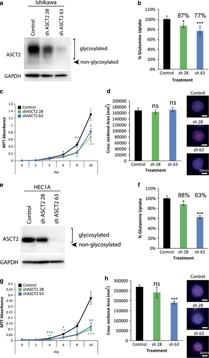
Figure 6.
shRNA knockdown of ASCT2 largely recapitulates the results of chemical inhibition. (a) Western blot of shASCT2 28 (sh 28)- and 63 (sh 63)-mediated knockdown of ASCT2 in Ishikawa cells. (b) The effect of ASCT2 sh 28 and sh 63 on [3H]-l-glutamine uptake in Ishikawa cells. (c) MTT cell viability assays showing the effect of ASCT2 sh 28 and sh 63 on Ishikawa cells. (d) 3D spheroid cross-sectional area of Ishikawa cells expressing ASCT2 sh 28 and sh 63. (e) Western blot of shASCT2 28 and 63 mediated knockdown of ASCT2 in HEC1A cells. (f) The effect of ASCT2 sh 28 and sh 63 on [3H]-l-glutamine uptake in HEC1A cells. (g) MTT cell viability assays showing the effect of ASCT2 sh 28 and 63 on HEC1A cells. (h) 3D spheroid cross-sectional area of HEC1A cells expressing ASCT2 sh 28 and sh 63. Student’s t-test: *P<0.05, **P<0.01, ***P<0.001, NS P>0.05. Scale bar represents 20 μm.

