
Fig. 1.
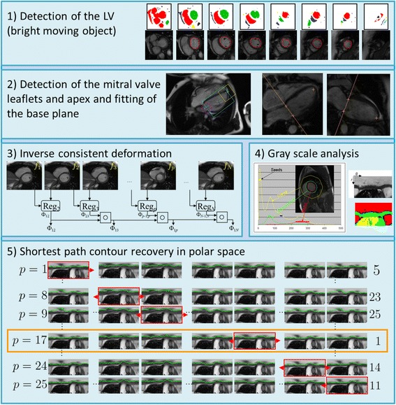
Automatic algorithm to segment the LV blood pool cavity and myocardium. Each box shows a step in the algorithm. 1) Detection of the blood pool as the bright moving object. 2) Detection of the mitral valve anchor points in the long axis images and fitting of the mitral valve base plane over time. 3) Registration of neighboring frames to establish deformation fields between the first frame and any other frame in the time series. 4) Histogram analysis to determine the major regions in the image, namely, blood, myocardium, and lungs. 5) Shortest path algorithm to recover a contour, endocardium or epicardium in polar space