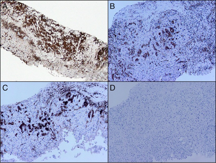
Figure 3.

Immunohistochemical stains on liver biopsy. (A) CD163 showing numerous positive histiocytes filling the liver sinusoids (100x). (B) CD68 showing positivity in the histiocytes (100x). (C) Factor XIIIa showing weak to strong positivity in the histiocytes (100x). (D) CD1a is negative in the histiocytes (100x).
