Figure 3.
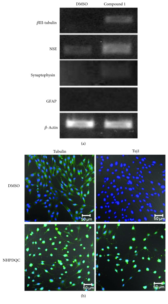
Neuronal differentiation of MSCs after treatment of NHPDQC. (a) Reverse transcription polymerase chain reaction (RT-PCR) results (left column: DMSO-treated group, right column: NHPDQC treatment group). Expression of the neuronal markers Tuj1 and NSE was increased in the MSCs at 48 h after treatment with NHPDQC. The RT-PCR results revealed that the expression of the presynaptic vesicle protein, synaptophysin, was increased slightly after 48 h of treatment with NHPDQC. Glial marker GFAP did not increase in both of undifferentiated and differentiated MSCs. (b) Immunofluorescence staining was performed in the DMSO-treated cell group and the group treated with NHPDQC. The results showed that Tuj1 expression was increased in the NHPDQC-treated group within 48 h. Green color: β-tubulin (upper panel), Tuj1 (lower panel); blue color: DAPI.

