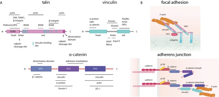
FIGURE 2:
Tension-sensitive proteins are mechanical players of adhesion sites. (A) Schematic representation of the main tension-sensitive proteins involved in focal adhesions and adherens junctions: talin (purple), vinculin (light blue), and α-catenin (dark blue). The main protein interaction domains are shown, and the known interactors (with their binding sites) are indicated above or below each protein. (B) Top, components of focal adhesions and the structures of talin and vinculin when stretched (talin in pink, vinculin in light blue, PIP2 in purple, and F-actin in orange). Bottom, components of adherens junctions and the structure of the unstretched (closed, top) or stretched (bottom) α-catenin (cadherin in purple, p120 in yellow, β-catenin in coral, α-catenin in purple, vinculin in light blue, and actin in orange).

