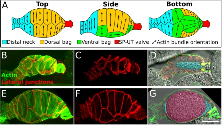
FIGURE 1:
Anatomy of the C. elegans spermatheca. (A) Schematic diagram showing actin bundle orientation in spermathecal cells. (B–G) Confocal images of two fixed and stained spermathecae, one that is unoccupied, sperm only (B–D), and one that is occupied, sperm and oocyte present (E–G). (B, C, E, F) Confocal maximum intensity projections of spermathecae expressing INX-12::mApple to label lateral junctions (red) stained with phalloidin to label F-actin (green). Note the difference in cell stretch in an unoccupied (C) and an occupied (F) spermatheca. (D, G) A central sagittal z-slice showing a cross section of the spermatheca with basal actin bundles (green), lateral junctions (red), and bright-field image (grayscale). Sperm and oocyte are false colored in blue and pink, respectively. Scale bar, 20 μm. In all images, the spermatheca is oriented distal to proximal.

