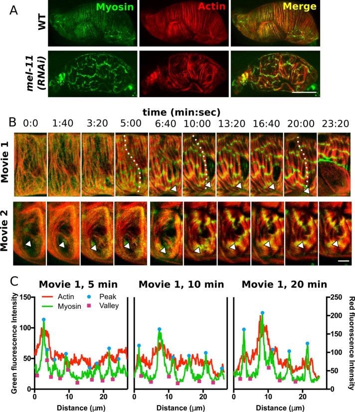
FIGURE 9:
Elevated myosin activity alters myosin organization and interaction with actomyosin bundles. (A) Confocal maximum intensity projections of fixed spermathecae from a WT animal and an animal treated with RNAi against myosin phosphatase, mel-11(RNAi), expressing moeABD::mCherry to label actin (red) and GFP::NMY-1 to label myosin (green). Note the homogeneous distribution of myosin in actomyosin bundles in WT and laterally associated myosin clusters that create transverse bands across several actomyosin bundles in a mel-11(RNAi) spermatheca. (B) Confocal maximum intensity projections from two 4D ovulation movies showing progression of mel-11(RNAi) phenotypes in selected cells. In movie 1, the actomyosin bundles rupture. The arrowhead indicates the site of bundle detachment. At 6:40, bundles are attached, and they retract by 10:00. By 23:20, the basal cell layer pulls back (last frame). In movie 2, misaligned actin bundles appear to be stabilized and reinforced (arrowhead). (C) Line-scan analysis along a single actomyosin bundle in movie 1 (indicated by a dashed line) at 5, 10, and 20 min after oocyte entry reveals that myosin clusters increase in intensity and grow by fusion of smaller clusters along a single bundle. In addition, actin fluorescence intensity is increased under prominent myosin peaks. Scale bar, 20 μm (A), 5 μm (B).

