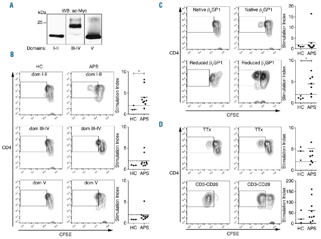
Figure 1.
Domain I–II and reduced β2GP1 induce proliferation of T cells from APS patients. (A) β2GP1 recombinant domains are purified and analyzed by Western blot. Data are representative of 3 independent experiments. (B–D) Flow cytometry analysis of the proliferation assay performed on PBMC of APS patients in the presence of domains I–II, II–IV, V β2GP1, reduced β2GP1 and tetanus toxin (TTx). CD3-CD28 is positive proliferation control. CD4+CFSElow population is gated on CD3+ cells. Data are mean ± SEM of 9 different donors. Statistical significance was determined by Mann-Whitney U analysis. HC: healthy controls; APS: Antiphospholipid syndrome; WB: western blot; ac-MYC: anti-c-Myc; CFSE: carboxyfluorescein diacetate succinimidyl ester.

