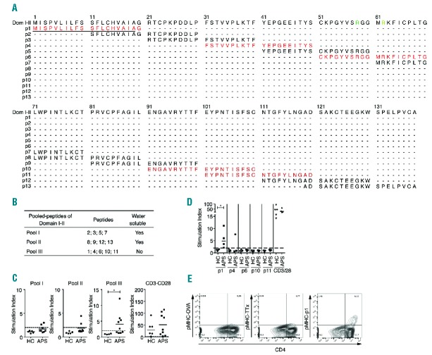
Figure 2.
Peptide 1 of β2GP1 carries antigenic determinant for CD4+ T-cell proliferation. (A) Representation of the peptide library of domain I–II. Peptides in red are peptides contained in Pool III. Residues in green represent R39-R43 epitopes. Underlining represents a signal peptide. (B) Table of peptide distribution for proliferation assay. (C) Flow cytometry analysis of proliferation assay performed on PBMC of APS patients in the presence of pool I, II or III. CD3-CD28 shows positive proliferation control. (D) Flow cytometry analysis of proliferation assay performed on the PBMC of APS patients in the presence of p1, p4, p6, p10 and p11. CD3-CD28 is positive proliferation control. CD4+CFSElow population is gated on CD3+ cells. Data are mean ± SEM of 9 different donors. Statistical significance was determined by Mann-Whitney U analysis. (E) Flow cytometry analysis of pMHC class II tetramer staining loaded with p1 on PBMS of APS patients pulsed with recombinant domain I–II. pMHC-OVA (ovalbumin) and pMHC-TTx (Tetanus toxin) are negative controls. Populations are gated on CD3+ cells. HC: healthy controls; APS: antiphospholipid syndrome.

