Figure 2.

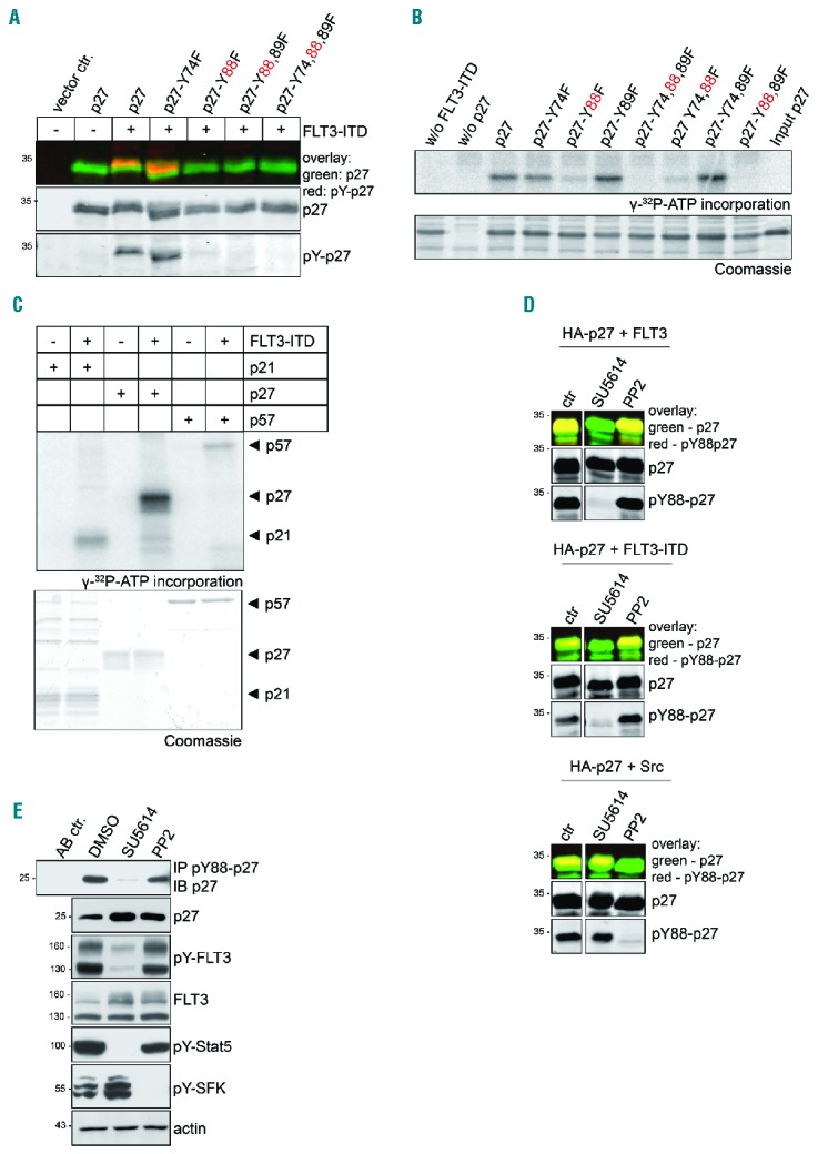
FLT3 phosphorylates p27 selectively on Y88 in vivo and in vitro and Y88-phosphorylation of p27 is Src family tyrosine kinase (SFK) independent. (A) FLT3-ITD phosphorylates p27 on Y88. In a similar approach as described in Figure 1G, HA-p27 and HA-p27 mutants where tyrosines were replaced by phenylalanine were expressed together with FLT3-ITD in 293T cells. p27 was detected simultaneously with overall tyrosine phosphorylation in heat-treated extracts. Co-migration indicates that the tyrosine-phosphorylated protein is p27 (pY-p27). (B) FLT3-ITD phosphorylates p27 on Y88 in vitro. Kinase assays were performed with recombinant His-p27 or His-p27 mutant proteins as indicated. The Flag-tagged cytoplasmic domain of FLT3-ITD was expressed in 293T cells and precipitated with anti-Flag antibodies. p27 protein or mutants were incubated with the sepharose-A bound kinase and γ-32P-ATP. Incorporation of γ-32P-ATP in p27 was detected by autoradiography (top panel) following SDS-PAGE and Coomassie brilliant blue staining (bottom panel). (C) FLT3-ITD selectively phosphorylates p27. In vitro kinase assay was performed as described above (B) using the cytoplasmic domain of recombinant FLT3-ITD isolated from insect cells as kinase and recombinant His-p21, His-p27 and His-p57 as substrates. (D) The receptor tyrosine kinase inhibitor SU5614, but not the SFK inhibitor PP2, decreases pY88-p27 after FLT3 or FLT3-ITD expression. 293T cells were treated with SU5614 (1 μM), PP2 (10 μM), or vehicle (DMSO) for 3 hours (h) following transient tansfection of HA-p27 together with FLT3 (top panel), FLT3-ITD (middle panel), or Src (bottom panel). Western blot analysis was performed as described in Figure 1G. (E) p27-Y88 phosphorylation occurs independently of activated SFK members. 32D-FLT3-ITD cells were treated with SU5614 (1 μM), PP2 (10 μM), or vehicle (DMSO) for 2 h. pY88-p27 was immunoprecipitated with pY88-p27-specific antibody and analyzed by Western blot with HRP-coupled anti-p27 antibodies. pY694-Stat5 serves as a control for SU5614 and pY416-SFK for PP2 treatment. Actin was used as loading control. One representative experiment of three independent experiments is shown. ctr: control (vehicle-treated cells).
