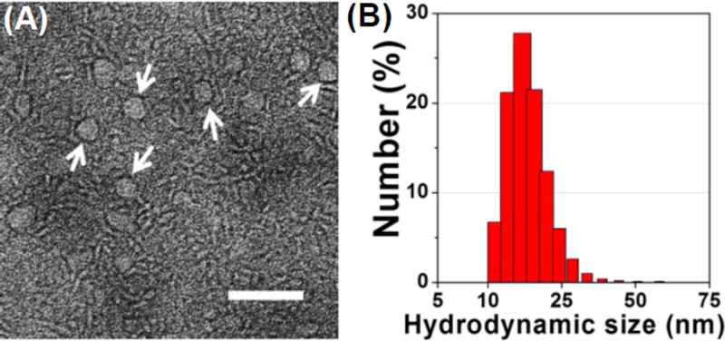
Fig. 3.
Characterization of Zol-Ca@bi-lipid NPs prepared with a Zol-Ca@DOPA to DSPE-PEG2k ratio of 20 µg/1 mg. (A) a representative TEM image after negative staining. Nanoparticles with clear core-shell structure are indicated by white arrows (bar = 100 nm); (B) hydrodynamic size distribution of the Zol-Ca@bi-lipid NPs in aqueous suspension.

