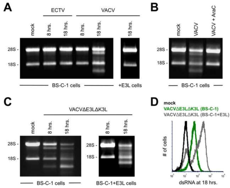
Figure 6. RNase L activation is observed only in VACV-infected cells at late time points.
(A) BS-C-1 or BS-C-1+E3L cells were infected (MOI=5) with either ECTV or VACV. Total RNA was isolated at the indicated time points and separated using a 1% bleach TBE gel. Each lane contained 4 µg of RNA. The data are representative of three independent trials. (B) BS-C-1 cells were infected (MOI=5) with VACV either in the presence of absence of AraC. RNA breakdown was detected as above at 18 hrs. post-infection. (C) BS-C-1 or BS-C-1+E3L cells were infected (MOI=5) with VACVΔE3LΔK3L. RNA breakdown was detected as above at the indicated time points. (D) BS-C-1 or BS-C-1+E3L cells were infected (MOI=10) with VACVΔE3LΔK3L for 18 hrs. prior to staining for dsRNA. The flow cytometric data are depicted in histogram format and representative of two independent trials.

