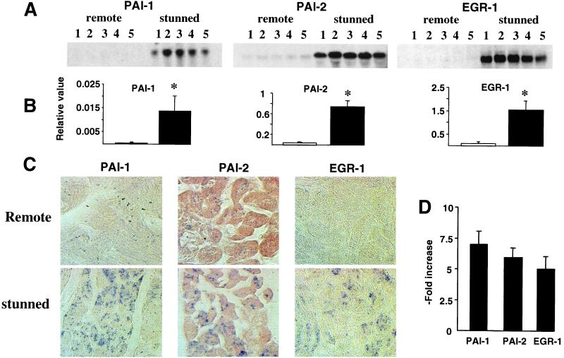
Figure 2.
Changes in gene expression analyzed by Northern blotting and in situ hybridization. (A) Northern blot was performed on samples of the remote and stunned area from five different hearts at 1-h reperfusion. Transcripts coding for PAI-1, PAI-2, and EGR-1 are illustrated. (B) Differences after 18S rRNA normalization between remote myocardium (open bars) and stunned myocardium (closed bars). *, P < 0.05 versus remote. (C) Tissue distribution by in situ hybridization. A clear induction was observed in cardiomyocytes from the ischemic area (stunned). (D) Changes in gene expression quantitated in isolated cardiomyocytes, prepared from the remote and stunned area of three different hearts submitted to 90-min ischemia and 1-h reperfusion. The graph illustrates the n-fold increase of PAI-1, PAI-2, and EGR-1 transcripts in stunned over normal myocytes.

