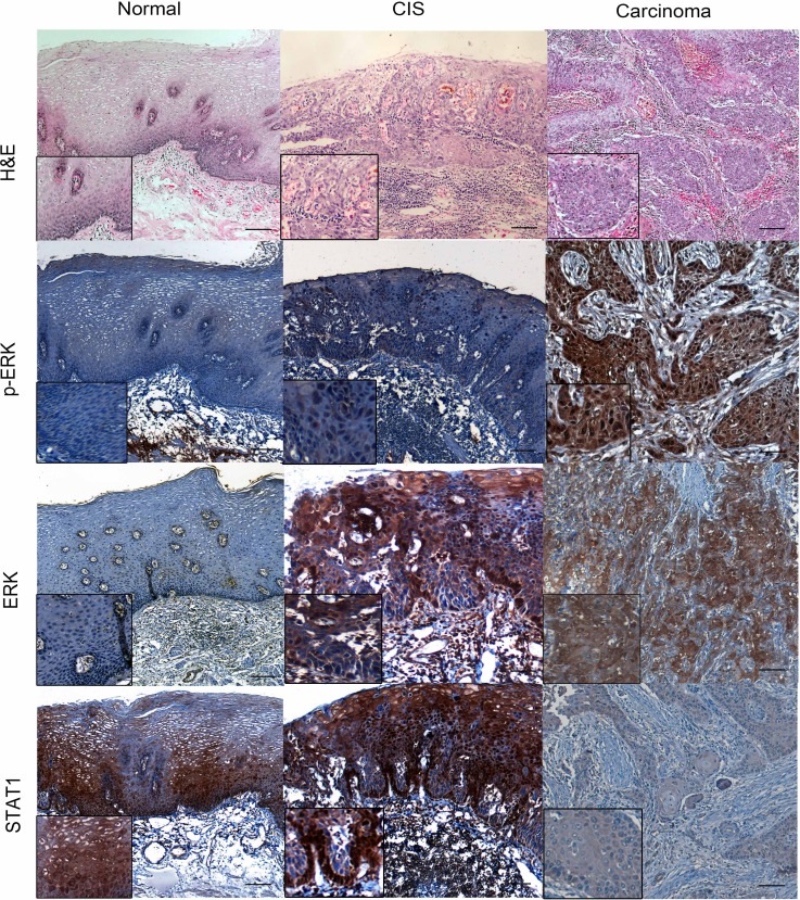
Figure 2. p-ERK, ERK and STAT1 expression in normal epithelial, carcinoma in situ (CIS) and ESCC patient samples.
The esophageal normal epithelial, CIS and carcinoma tissues were stain by Hematoxylin and eosin (H&E). By immunohistochemistry applied to formalin-fixed paraffin-embedded tissues, variable levels of p-ERK, ERK and STAT1 were detected in normal esophageal, CIS and carcinoma examined. (IHC stain, scale bar, 50 μM).

