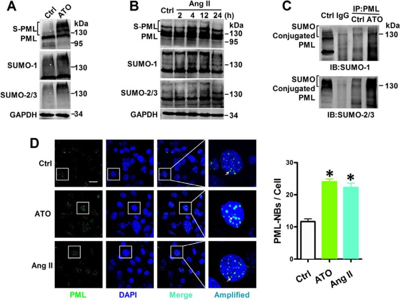
Figure 2. Effects of ATO and Ang II on PML SUMOylation and PML-NBs formation.
Whole cell extract were treated with anti-PML, anti-SUMO-1 and anti-SUMO-2/3 antibody in NMCMs that were exposed to (A) ATO (2 μM) for 2 h and (B) Ang II (100 nM) for the indicated periods. The protein molecular masses are in kDa and shown to the right of each panel. The high molecular-mass-weight bands (> 130 kDa) in parentheses represent SUMOylated PML (S-PML). (C) Total lysates from NMCMs were immunoprecipitated with anti-PML antibody. The immunopellets then were detected with either anti-SUMO-1 or anti-SUMO-2/3 antibody. (D) Confocal immunofluorescent analysis of PML-NBs (green) and nuclei (blue) in NMCMs treated with ATO (2 μM) for 2 h or Ang II (100 nM) for 4 h. Scale bar: 20 μm. Enlarged view of a single PML-NB in the boxed region is shown at higher magnification in the right panel. White arrowheads indicate the typical PML-NBs. *P < 0.05 versus control group. The data shown in Figure A–D are representative of three separate experiments.

