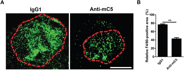
Figure 4. Anti-C5 antibody prevents infiltration of F4/80-positive cells in CNV lesions.

(A) Representative photographs of F4/80-positive cells in CNV lesions at 3 days after laser photocoagulation and intravitreal injection of anti-C5 antibody. Red dashed lines delineate CNV areas. Scale bar, 200 μm. (B) Quantitative analyses of relative F4/80-positive areas of CNV lesions (n = 6). Anti-mC5, anti-C5 antibody; IgG1, IgG1 isotype control. **, P-value < 0.01 (Mann-Whitney U-test).
