
Figure 5. PES and PET-16 alter HSP70 ATPase activity and mitochondrial protein quality control.
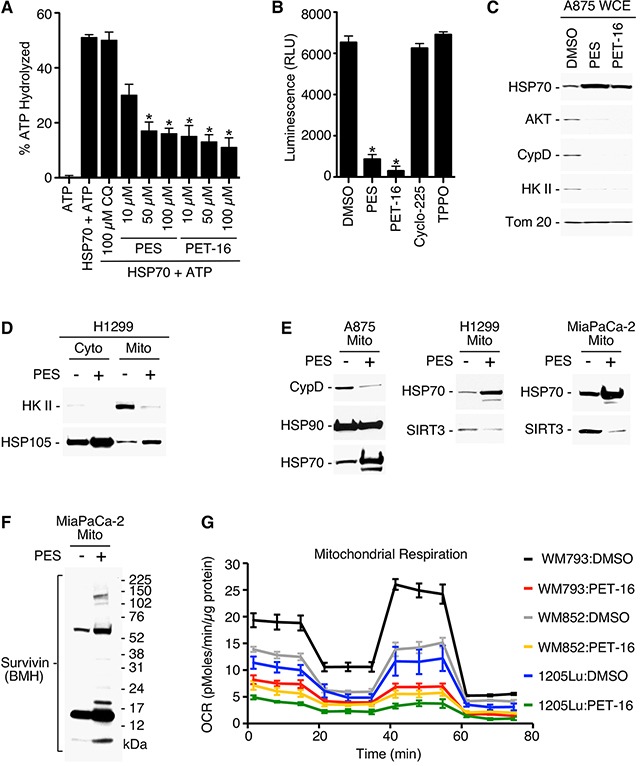
(A) Purified full-length HSP70 protein generated in bacteria was incubated with each of the compounds specified and assayed for intrinsic ATPase activity. Results shown are the mean ± SD of at least three independent experiments. *P<0.05. (B) A875 human melanoma cell line stably expressing a luciferase expression construct was treated with the indicated compounds and assayed for relative luciferase luminescence (RLU). Each graphical representation indicates the mean ± SD of at least three independent cultures relative to control cells. *P<0.01. (C) A875 cells were treated with 20 μM PES or PET-16 as indicated. Cell lysates were fractionated into detergent-soluble preparations and assayed by Western blotting for the proteins indicated. (D-F) The cell lines indicated were treated with PES, mitochondrial and/or cytosolic fractions were isolated, and western blots probed for the proteins indicated. (G) Mitochondrial oxygen consumption rate (OCR) in the melanoma cell lines indicated was measured before and after treatment with 1 μM PET-16 for 24 h. Each graphical representation indicates the mean ± SD of at least three independent cultures.