
Figure 6. ERBB4 expression regulates EMT in MNK45 cells induced by TGF-β1.
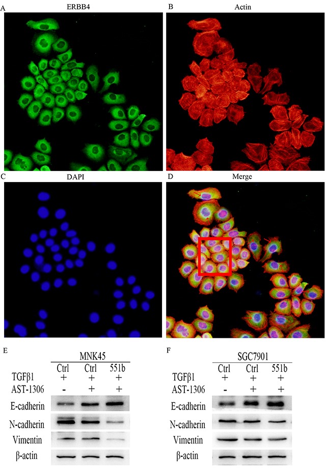
(A) Immunoflorescence staining with anti-ERBB4 antibody. (B) Cytoskeleton staining: of MNK45 cells induced by TGF-β1 were stained by TRITC conjugated Phalloidin. (C) Nuclear DAPI staining of GC cells induced by TGF-β1. (D) Merge of A-C Red arrows point to EMT cells showing stretched out lamellipodia and filopodia and enhanced cytoplasmic expression of ERBB4. The red box shows cells with low ERBB4 expression with non-EMT characteristics. The images shown are under 200x magnification. (E and F) Inhibition of EMT due to ERBB4 inhibition by AST-1306 in MNK45 and SGC7901 cells by Western blot analysis of E-Cadherin, N-Cadherin and Vimentin are shown. β-actin was used as internal control.