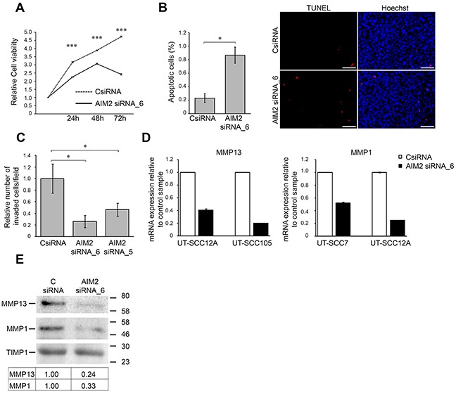
Figure 4. Knockdown of AIM2 inhibits viability and invasion of cSCC cells.

(A) The number of viable cSCC cells (UT-SCC7) was determined 24, 48, and 72 hours after transfection with AIM2 siRNA_6 and control siRNA (CSiRNA) (75 nM) (mean ± SD, n=8). (B) cSCC cells (UT-SCC7) were transfected with AIM2 siRNA_6, or control siRNA and 48 hours after transfection apoptotic cells were detected with TUNEL staining and the relative number of TUNEL positive apoptotic cells with positive nuclear staining was determined by counting 2000-3000 cells at 10× magnification per image (n=3, mean ± SD) (left panel). Representative images of the quantification are shown. Scale bar=100 μm (right panel). (C) UT-SCC7 cell lines were transfected with AIM2 or control siRNAs and seeded to the inserts coated with type I collagen 24 hours after transfection. After 48 hours the number of invaded cells was counted (mean ± SD, n=3). (D) MMP13 and MMP1 mRNA levels were determined by qRT-PCR 72 hours after AIM2 knockdown in cSCC cells. (E) Levels of MMP13 and MMP1 in conditioned media of cSCC cells (UT-SCC12A) were determined by Western blot analysis 72 hours after AIM2 knockdown. TIMP1 was used as a marker for equal loading. Levels of MMP1 and MMP13 were quantitated by densitometry and corrected for the level of TIMP1. (*P<0.05, ***P<0.001; Student's t-test).
