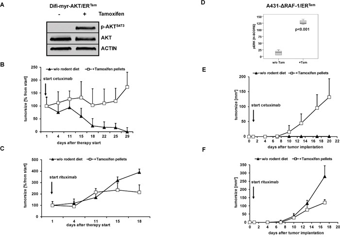
Figure 4. Activated PI3K/AKT and MAPK signaling protects tumors against anti-EGFR antibody-mediated cytotoxicity in vivo.

(A) Palpable flank tumors were established by subcutaneous injection of A431-myr-AKT/ERTam cells in NOD/SCID mice. Tumor-bearing mice were fed with diet with or without tamoxifen for transgene activation in vivo for one week. A strong phosphorylation of AKT as marker of PI3K/AKT signaling was detected in protein lysates from explanted tumors by immunoblot analyses. (B, C) Tumor growth following injection of Difi-myr-AKT/ERTam cells in NOD/SCID mice fed with (open boxes) or without (closed triangles) tamoxifen. After tumors were palpable (arrow), mice were treated biweekly with intraperitoneal injections of cetuximab (0.5 mg) (B) or rituximab (1 mg) (C). Mean bidimensional tumor sizes (+ SD) of 5 mice per group are given. (E, F) NOD/SCID mice were fed with diet with or without tamoxifen for one week before A431-myr-ΔRAF-1/ERTam cells were subcutaneously implanted. The day after the tumor implantation mice were treated biweekly with intraperitoneal injections of cetuximab (1 mg) (E) or rituximab (1 mg) (F). Tumor growth in NOD/SCID mice fed with (open boxes) or without (closed triangles) tamoxifen was measured bidimensional twice weekly. Mean bidimensional tumor sizes (+ SD) of 5 mice per group are given. (D) Palpable flank tumors of mice treated with rituximab were explanted and analyzed by immunhistochemistry. A strong phosphorylation of ERK1/2 was detected in explanted tumors of mice fed with tamoxifen diet.
