
Figure 8. IL-12 and IL-18 synergistically induce sustained phosphorylation of STAT4 and NF-κB p65 in human Vγ9Vδ2 T cells.
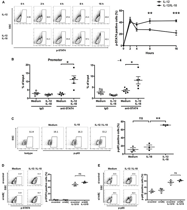
(A) Ex vivo-expanded Vγ9Vδ2 T cells were stimulated with IL-12 alone or in combination with IL-18 for different periods. Representative contour plots and percentage of p-STAT4 positive cells. The data for healthy donors (n = 4) was obtained from independent experiments. Bars represent the mean and SEM, **P < 0.01, ***P < 0.001, two-way ANOVA, followed by Sidak's multiple comparisons test. (B) Ex vivo-expanded Vγ9Vδ2 T cells were activated with IL-12 and IL-18 for 16 h followed by chromatin immunoprecipitation using anti-STAT4 Ab. Recruitment of STAT4 to the promoter (left) and −4 kb (right) of IFN-γ gene regions were analyzed by qPCR. The data were obtained from five independent experiments each using Vγ9Vδ2 T cells from different donors. Bars represent the mean and SEM, *P < 0.05, two-tailed paired Student t test. (C) Ex vivo-expanded Vγ9Vδ2 T cells were stimulated with IL-18 alone or in combination with IL-12 for 16 h. Representative contour plots and percentage of p-p65 positive cells. The data for healthy donors (n = 3) was obtained from independent experiments. Bars represent the mean and SEM, **P < 0.01, one-way ANOVA, followed by Tukey's multiple comparison test. (D, E) Ex vivo-expanded Vγ9Vδ2 T cells were transfected with siRNA targeting IκBζ or control siRNA followed by 16-hour treatment of IL-12 and IL-18. Representative contour plots and percentage of p-STAT4- (D) and p-p65- (E) positive cells by CD3+Vδ2 TCR+ cells. The data were obtained from three independent experiments each using Vγ9Vδ2 T cells from different donors. Bars represent the mean and SEM, two-tailed paired Student t test.