
Figure 3. The induction of nuclear DNA double-strand break by DHA in Cal-27 cells.
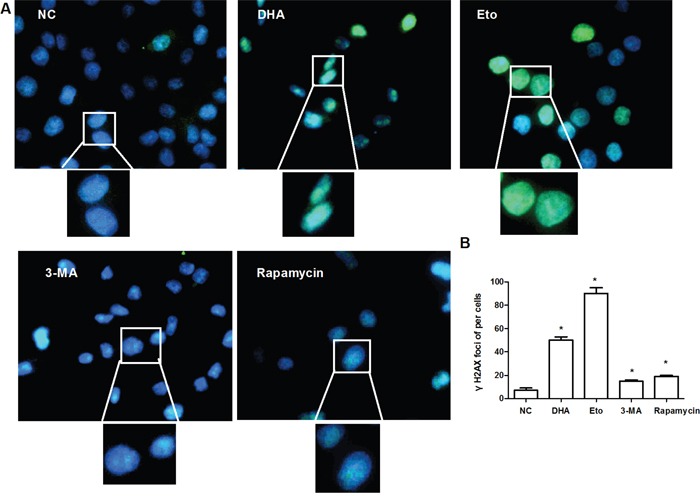
(A) Representative images of γH2AX foci formation. Cal-27 cells were treated with 0.1% DMSO or DHA (24.5 μM) for 24 h, and analyzed for γH2AX (green). Etopside (40μM) was used as DNA DSB positive control. 3-MA (1 mM) and rapamycin (0.1 μM) acted as autophagy inhibitor and activator, respectively. Nuclei were counter-stained with DAPI (blue). The upper and bottom panels are respectively 400× and 1000×. (B) Statistical analysis of the number of γH2AX foci. Data are shown as the mean ± SD (n = 3). * P < 0.05 vs. NC group.