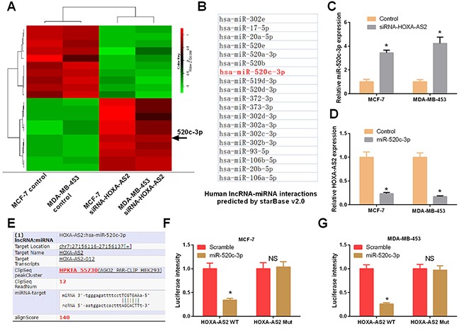
Figure 5. Mir-520c-3p was directly regulated by HOXA-AS2 in breast cancer.

(A) MCF-7 and MDA-MB-453 cells were transfected with HOXA-AS2 siRNAs or control for 48 hrs. Hierarchical clustering revealed systematic variations in the expression of miRNAs. Numerous differentially expressed miRNAs between control and HOXA-AS2 siRNAs transfected MCF-7 and MDA-MB-453 cells are shown on a scale from green (low) to red (high). The arrow indicates miR-520c-3p, which was included in these miRNAs. (B) StarBase v2.0 predicted the HOXA-AS2-regulated miRNAs (red one was miR-520c-3p). (C) qRT-PCR analysis of miR-520c-3p expression in MCF-7 and MDA-MB-453 cells transfected with HOXA-AS2 siRNAs or control (*P < 0.05). (D) qRT-PCR analysis of HOXA-AS2 expression in MCF-7 and MDA-MB-453 cells transfected with miR-520c-3p mimics or control (*P < 0.05). (E) StarBase v2.0 results showing the sequence of HOXA-AS2 with highly conserved putative miR-520c-3p binding sites. (F, G) MCF-7 and MDA-MB-453 cells were co-transfected with the reporter plasmid (or the corresponding mutant reporter) and miR-520c-3p. The relative fluorescence value was detected by luciferase reporter gene assays (*P < 0.05).
