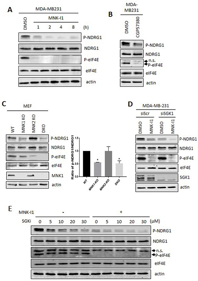
Figure 2. Pharmacological and genetic evidence that MNKs modulate the phosphorylation of NDRG1.

(A) MDA-MB-231 cells were treated with 5 μM MNK-I1 for the indicated times and lysates were then analysed by Western blot. (B) MDA-MB-231 cells were treated with 25 μM CGP57380 for 2 h. Lysates were analysed by Western blot. (C) Lysates from wild type, MNK1-KO, MNK2-KO, and MNK1+MNK2 double knockout (DKO) MEFs were analysed by immunoblot with the indicated antibodies. Ratio of p-NDRG1/NDRG1 was calculated using Image J. Data are shown as mean ± S.E.M. from three replicates. *P < 0.05. (D) SGK1 was knocked down by an siRNA in MDA-MB-231 cells and, where shown, cells were then treated with 5 μM MNK-I1 for 4 h. Lysates were analysed by Western blot. (E) MDA-MB-231 cells were treated with increasing concentrations of SGK inhibitor (SGKi) together with 5 μM MNK-I1, where indicated, for 4h. Lysates were analysed by Western blot. In some cases, the antibody used for P-eIF4E recognises an additional band which appears to be a non-specific reaction and is not affected by the MNK inhibitors (indicated as ‘n.s.’ with an arrowhead in panels B and E).
