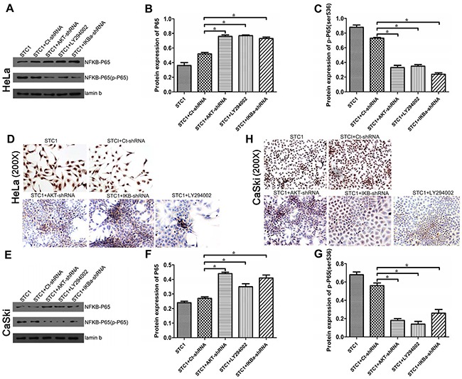
Figure 5. The nucleprotein and phosphorylation levels of P65/phospho-P65 (Ser536) in cervical cancer cells.

NF-κB P65 nucleprotein in STC1 overexpressed HeLa (A, B) and CaSki (E, F) cell lines treated with the AKT-shRNA, PI3K inhibitor LY294002 and IκBα-shRNA were detected by Western blotting. Phospho-P65 (Ser536) nucleprotein in STC1 overexpressed HeLa (C, D) and CaSki (G, H) cell lines treated with the AKT-shRNA, PI3K inhibitor LY294002 and IκBα-shRNA were detected by Western blotting and ICC. n=3, *p<0.05.
